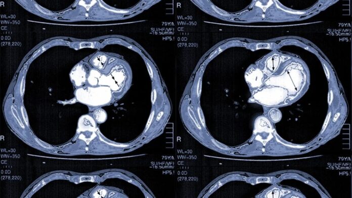
Cardiologists play a fundamental role in managing cardiovascular health. They use their expertise to diagnose, treat, and prevent heart diseases, which remain a leading cause of mortality worldwide. With advancements in medical technology, cardiologists now rely on imaging tools to gain precise insights into the health of their patients’ hearts. These technologies are key in detecting conditions early and helping patients lead healthier lives. Here’s information on the primary cardiology imaging tools cardiologists use to diagnose heart conditions effectively:
Echocardiograms
Heart disease refers to various heart-related conditions, such as coronary artery disease, arrhythmias, and heart valve problems. Early diagnosis is fundamental, and cardiology imaging serves as a foundational element in achieving this. An echocardiogram, or ECHO, is a noninvasive imaging tool that uses high-frequency sound waves to create detailed images of the heart. By offering a real-time view of the heart’s chambers, valves, and blood flow, ECHO is one of the most relied-upon methods in cardiology.
This tool is painless and safe, making it a first-line option for many diagnostic scenarios. Some key uses of ECHOs are:
- Detecting structural abnormalities, such as valve disorders or congenital defects.
- Monitoring the effectiveness of treatments for various heart conditions.
- Assessing overall heart function, including identifying weakened heart muscles.
Electrocardiograms
An electrocardiogram, or ECG/EKG, measures the electrical activity of the heart to determine irregularities in its rhythm and overall function. While not a traditional imaging test, it provides significant diagnostic insights. It helps detect arrhythmias such as atrial fibrillation, determine signs of a past or impending heart attack, and assist in evaluating the causes of chest pain or unexplained fatigue. This quick and noninvasive test is a fundamental diagnostic tool for both clinical evaluations and emergency scenarios.
Ultrasounds
Cardiac ultrasounds, or ultrasounds targeting the cardiovascular system, provide another layer of insight into heart health. This tool evaluates blood flow and assesses the vessels that carry it to and from the heart. Cardiac ultrasounds are used to detect plaque buildup in arteries, assess the severity of peripheral artery disease, and analyze structural issues in arteries and veins that may affect cardiac performance. Like echocardiograms, cardiac ultrasounds are noninvasive, efficient, and provide results that guide treatment decisions.
Coronary Angiography
Coronary angiography is an advanced imaging technique that offers a detailed look at the heart’s blood vessels. This minimally invasive test uses a special dye visible on X-rays, combined with a catheter inserted into the coronary arteries. While more invasive than other diagnostic methods, coronary angiography is indispensable in evaluating coronary artery disease and planning interventions. Some key uses of coronary angiographies include:
- Identifying blockages or narrowing of the coronary arteries.
- Pinpointing the causes of chest pain or abnormal stress test results.
- Helping cardiologists formulate personalized treatment plans, such as angioplasty or stenting, in cases of significant blockages.
Learn How Cardiology Can Impact Your Heart Health
Cardiology imaging tools play a pivotal role in diagnosing and managing heart diseases. From the precision of echocardiograms to the insights gained through coronary angiography, these technologies provide cardiologists with the information they need to safeguard your heart health. To take charge of your cardiovascular well-being, schedule a consultation with an experienced cardiologist today.

